
-
View Larger Image
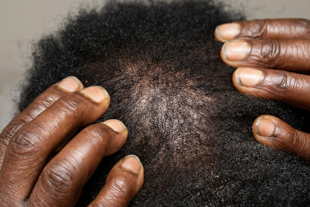
Can Phone Photos Reliably Measure a Widening Hair Part?
Hair monitoring and measurement
Can Phone Photos Reliably Measure a Widening Hair Part?
Phone photos can help document change when position, lighting, hair preparation, camera settings, and scale are repeated. A casual photo cannot diagnose hair loss, and a changing-looking part may reflect styling, moisture, color, angle, or exposure rather than a true density change.
Direct answer: A series of carefully standardized phone photos can show whether a part appears different over time, but it is not a reliable standalone measurement unless the same part line, hairstyle, lighting, camera geometry, scalp condition, and scale are reproduced. Even a convincing visual change does not identify the cause.
This guide is for consumers, salon professionals, and anyone helping document a gradual change. It explains what photographs can contribute, why informal comparisons mislead, and when a widening part deserves medical evaluation.
Why a hair part can look wider without true density loss
A hair part is a styled line through a three-dimensional field of fibers. Its apparent width changes when the comb follows a slightly different route, the head tilts, the camera moves, or hair lies flatter. Wetness, oil, conditioner, static, product residue, new gray fibers, root color contrast, and curl shrinkage can reveal more or less scalp. Flash and overhead light can brighten the scalp and darken adjacent hair, exaggerating contrast.
That does not make photographs useless. It means the photograph is a measurement system with variables that must be controlled. Researchers and clinicians have long emphasized consistent position, lighting, magnification, styling, and views when tracking hair. A 2024 practical guide developed standardized protocols for different hair-loss patterns and concluded that consistent photography improves objective clinical assessment. Review the PubMed record.
What standardized photography can do
Standardized global photos can preserve a visual baseline, help identify the distribution of apparent thinning, and support a conversation with a clinician. They can also show whether a concern is confined to one part line or appears across the vertex, temples, frontal hairline, or patches.
A 2025 quality-improvement study in an academic dermatology clinic trained staff to obtain consistent minimum views. Correct standardized photo collection rose from 32 percent in the initial sample to 66 percent after the intervention. The study supports a practical point: training and a repeatable protocol improve documentation. It does not show that a phone photo alone diagnoses a condition or precisely counts follicles. Read the open-access study.
Image resolution matters differently for different questions. Research using standardized alopecia images found that clinicians could identify broad regions of loss at relatively modest resolution, while identifying vellus-like fibers required much higher detail. The investigators also showed that a styling part can be mistaken for lower density when contextual information is lost. See the image-resolution study.
Evidence limit: Photos are strongest for documenting visible distribution and change under reproduced conditions. They are weaker for measuring individual fiber diameter, miniaturization, follicle density, inflammation, scarring, or the cause of loss. Those questions may require examination, trichoscopy, laboratory assessment, or biopsy.
A practical home photo protocol
1. Choose a baseline day
Photograph clean, fully dry hair before fibers become heavily coated or compressed. Record when the hair was washed, whether conditioner or styling product was used, and whether color or chemical service recently changed contrast. Do not delay needed medical care just to obtain a perfect baseline.
2. Reproduce the part
Choose a clear landmark, such as the center of the nose to the crown, and use the same combing direction each time. Do not force a new narrow part to make the image look better or repeatedly widen it to search for a problem. Take one untouched view before adjusting the hair.
3. Fix the camera geometry
Use the same room, device, lens, orientation, distance, and head position. A helper or stable phone support is usually more repeatable than an arm-length selfie. Avoid digital zoom. Place the camera perpendicular to the scalp region rather than at an angle that shortens one side of the part.
4. Control the light
Use diffuse light from the same direction and avoid mixing bright window light with warm indoor bulbs. Turn flash either consistently on or consistently off. If scalp shine is strong, note that rather than editing it away.
5. Include a safe scale
A clean ruler or measurement marker can be placed on the same plane near the part without pressing hair aside. The scale helps with image calibration, but only when it is not closer to the camera than the scalp. Never place a shared unclean tool against irritated or broken skin.
6. Capture more than one view
Take an overhead vertex view, frontal hairline view, and left and right oblique views. Consistent multiple views provide context that a single close-up lacks. Keep the background simple and crop out unnecessary personal information.
7. Compare at useful intervals
Day-to-day comparisons amplify noise from washing, styling, humidity, and lighting. For a gradual concern, use an interval recommended by a clinician or a consistent monthly schedule. If change is sudden, patchy, painful, or inflammatory, seek evaluation rather than waiting for a photo series.
Can you measure the part in pixels or millimeters?
You can measure the visible light scalp band at predefined points when every image includes a same-plane scale and replicated geometry. But a number can create false confidence. The edge between hair and scalp is not always clear, fibers cross the part, and lighting changes the apparent boundary. Measuring three or more fixed points is more informative than selecting the widest point after seeing the photo.
Professional systems may use stereotactic positioning, trichoscopy, image segmentation, or a marked scalp area to improve reproducibility. A review of hair-evaluation methods notes that fixed camera and head positioning, repeated styling, and consistent views are central to meaningful comparison. Review the measurement-method overview.
What a widening part may mean
A widening central part is a recognized possible sign of patterned hair loss, especially when change is gradual and accompanied by reduced fullness. The American Academy of Dermatology lists a widening part among signs that may be noticed early. See the AAD overview.
It is not specific. Diffuse shedding, traction, breakage, inflammatory scalp disease, medication effects, illness, nutritional problems, hormonal changes, and styling can alter appearance. A clinician evaluates timing, distribution, symptoms, medical history, medications, family history, scalp findings, and hair-shaft condition rather than diagnosing from one picture.
When salon documentation is useful
Salon professionals can help clients create consistent, non-diagnostic records. Obtain consent, use neutral language, keep images secure, and document service variables that could change appearance. A useful note says, “The visible part appeared wider than in the prior standardized image under the same setup,” not “The client has androgenetic alopecia.”
Refer when the change is sudden, patchy, painful, burning, itchy, crusted, draining, or associated with shiny areas that appear to lack follicle openings. Also refer when eyebrows or body hair change, when shedding is heavy, or when the client is distressed or considering medication or supplements.
Common comparison mistakes
- Comparing wet hair with dry hair.
- Using flash in one image and diffuse light in another.
- Changing hair color, root coverage, or part placement.
- Moving the camera closer and treating the larger image as a wider part.
- Using beauty filters, sharpening, or automatic portrait blur.
- Comparing immediately after tight styling with a relaxed baseline.
- Measuring only the most concerning frame instead of predefined points.
Frequently asked questions
Can an app diagnose hair loss from a phone photo?
An app may organize images or estimate visible features, but performance depends on validated data, image quality, hair types represented, and the intended use. Do not treat a consumer estimate as a medical diagnosis.
Should I photograph shed hairs too?
A dated note about washing, brushing, and unusual shedding may help a clinician, but informal hair counts are affected by wash frequency and handling. Do not postpone evaluation to collect every strand.
Does a narrow part prove treatment is working?
No. Styling, fiber swelling, concealers, lighting, and camera position can narrow the visible scalp band. Treatment outcomes should be evaluated using appropriate clinical methods and realistic timelines.
Can a salon professional measure my part?
A salon professional can document observable width under a consistent protocol and refer concerns. Diagnosis, medication decisions, and treatment monitoring belong with appropriately licensed healthcare professionals.
The bottom line
Phone photos can make a gradual concern easier to describe when they are standardized. They cannot remove uncertainty or identify a cause by themselves. Reproduce the part, camera, light, scale, hair preparation, and interval; keep multiple views; avoid filters; and use the record to support, not replace, professional evaluation.
Explore related practical guidance in the Hair Society Tips & Tricks collection and learn more from the Hair Society hair-loss resources.
Share This Story, Choose Your Platform!
Related Posts
Can Scalp Massage Increase Hair Growth? What the Evidence Actually Shows
Gallery
Can Scalp Massage Increase Hair Growth? What the Evidence Actually Shows
Can Static Electricity Damage Hair or Mainly Change Manageability?
Gallery
Can Static Electricity Damage Hair or Mainly Change Manageability?

Does Swimming Pool Chlorine Damage Hair? What It Changes and How to Reduce Exposure
Gallery
Does Swimming Pool Chlorine Damage Hair? What It Changes and How to Reduce Exposure
Can Menopause Change Hair Texture as Well as Density?
Gallery
Can Menopause Change Hair Texture as Well as Density?